
Science
This New 3D Imaging Technique Depictures Blood Vessels
Here’s the VascuViz technique.

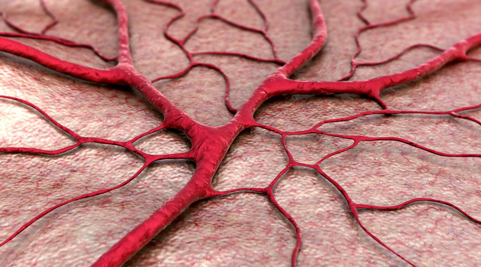
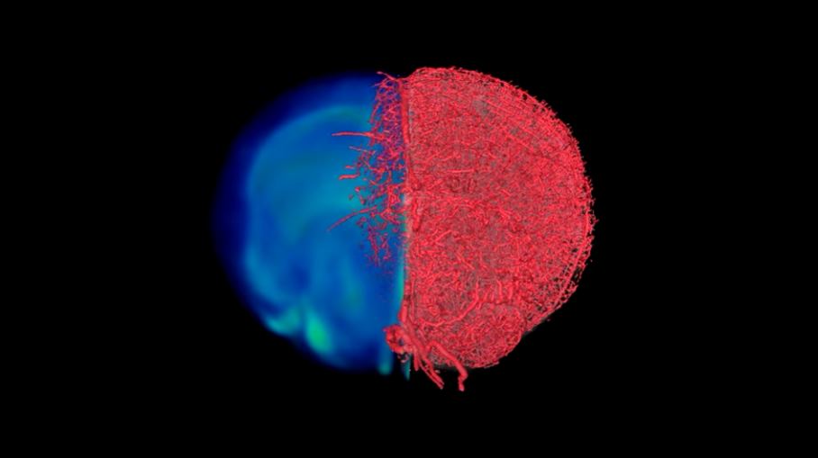
With the newly developed 3D visualization technique, the functioning of blood vessels is seen much more clearly. The technique, called VascuViz, uses a fast-curing polymer blend that makes blood vessels visible through a wide variety of scanning technologies as they move between tissues and organs.
So far, laboratory mouse tests show that it works at different scales, from the largest arteries to the smallest capillaries, and can be used to show details that would be missed by traditional techniques and improve our understanding of how tissues work.
New technique in researching blood vessels
“Now, instead of using an estimate, we can more precisely predict features like blood flow in real blood vessels and combine them with complementary information like cell density,” said mechanical engineer Akanksha Bhargava of the Johns Hopkins University School of Medicine.
With the measurements taken with VaskuViz, simulation models can be created that show how blood flow is, including cancer models, and a crucial advance can be made in understanding how such patients work.
What makes the new technique so useful is that it offers an all-in-one approach to results that would normally require several scans and several different methods to obtain.
“Usually, if you want to collect data about blood vessels in a particular tissue and combine it with all the surrounding context, such as the structure and the types of cells that grow there, you have to relabel the tissue several times, get multiple images and pieces of supplemental information together,” said Arvind Pathak, Associate Professor of Radiology and Oncology at the University of Medicine of John Hopkins. “This can be an expensive and time-consuming process that risks destroying tissue architecture and hampers our ability to use composite information in new ways.”
The VascuViz technique combines imaging modalities such as BriteVu (used in CT scans) and Galbumin-Rhodamine (used in MRI scans). What is thus produced is a wonderfully detailed, three-dimensional model of the blood vessel location.
The team behind the project said that cancer tumors, leg muscles, brain, kidney tissues, and the circulatory system can all be imaged by VascuViz. So the potential of this new approach is very high.
“We hope that these advances in preclinical vascular imaging, along with the new visualization approaches presented here, will open new perspectives for the image-based systems biology of the vasculature and help answer important questions in the broader field of microcirculation in health and disease,” the team wrote.
The research has been published in Nature Methods.

Mariana Trench: 8 Fascinating Facts About the Earth’s Deepest Place
10 most famous scientists who contributed to astronomy and space

Top 25 extremely stunning photos of Northern Lights of 2020 revealed

Meteorite makes Indonesian man a millionaire as piece crashes through his roof
It’s Now Possible to Predict Blood Cancer With Ten-Second Videos
This Inexpensive Method Speeds up Analysis of 3D Printed Metal Parts

SpaceX Dragon: A Game Changer for NASA
Tiny fish, big boom: This small fish found to roar like a gunshot

Turkish city unveils design of Mars Colony project for tourists

Researchers Reveal the Source of the Electron Rain Phenomenon
A New Study Reveals How Climate Change Will Affect Sound Travels Underwater
Here Are 6 Space Technologies We Can Benefit to Improve Life on Earth
SpaceX Announced Crew Dragon Will No Longer Be Produced
Russia Warns Sanctions on Moscow Could Endanger the ISS
A New Study Reveals How to Make Walking Easier

Remote-control cars to race on the Moon in 2021

Space Debris Problem Is Getting Worse: Causes and Removal Efforts of Rambling Junk
Microplastic Pollution Detected in Human Blood for the First Time

Nvidia CEO declares coding’s end in AI era

Here Is Blue Origin’s Touristic Rocket: The New Shepard Design
The Fastest Manned Plane in the World: Here’s The Insane Engineering of the X-15
Here Are 6 Space Technologies We Can Benefit to Improve Life on Earth
Pentagon Formed A New Group To Investigate UFO Reports
Space Debris Problem Is Getting Worse: Causes and Removal Efforts of Rambling Junk
From New Zealand to the US: The Rocket Lab Story
Here Is Blue Origin’s Touristic Rocket: The New Shepard Design
Take a Look at Earth From Space: Glasgow, Scotland

NASA Is Preparing Next Generation Spacesuits for Moon Mission and Beyond
Remote-control cars to race on the Moon in 2021
Meteorite makes Indonesian man a millionaire as piece crashes through his roof
NASA sends a new Space toilet for astronauts to ISS

 Earth & Energy5 years ago
Mariana Trench: 8 Fascinating Facts About the Earth’s Deepest Place

 Space6 years ago
10 most famous scientists who contributed to astronomy and space
- Skywatching6 years ago
Top 25 extremely stunning photos of Northern Lights of 2020 revealed
- Engineering6 years ago
Meteorite makes Indonesian man a millionaire as piece crashes through his roof

 Science5 years ago
It’s Now Possible to Predict Blood Cancer With Ten-Second Videos
 Engineering5 years ago
This Inexpensive Method Speeds up Analysis of 3D Printed Metal Parts
- Infographic6 years ago
SpaceX Dragon: A Game Changer for NASA

 Earth & Energy3 years ago
Tiny fish, big boom: This small fish found to roar like a gunshot